All You Need to Know About a Bone Density (DEXA) Scan
A bone density scan, or a DEXA scan, as the name implies measures the density of the bones. The scan measures the calcium and mineral content in the bones and reveals how strong the bones are. It helps to diagnose osteoporosis, or alternatively, ascertain a person’s risk for developing osteoporosis.
DEXA stands for dual energy x-ray absorptiometry. It is dual because two x-ray beams of different intensities are used. These beams are directed at the bones and the absorption of the beams is used to calculate the strength of the bones.
- DEXA scans can pick up even small variations in bone density. This helps to diagnose osteoporosis before something serious happens.
- DEXA scan is also used to check on the effectiveness of osteoporosis treatment.
- It helps to determine the composition of the body as well.
- It should be understood that a DEXA scan is not used to detect tumors or diagnose arthritis.
What Is Osteoporosis?
Our bones become thin as we age. Osteopenia is a condition in which the bones are thin and the bone mass is low. Low bone mass doesn’t necessarily translate to future osteoporosis. Quite a few athletes have low bone mass but they still have strong bones. Patients with osteopenia who also have other risk factors are more likely to develop osteoporosis.
Osteoporosis is an escalating condition in which the thinning of the bones continues till they become really fragile and prone to fractures. Osteoporosis literally translates to porous bones. While osteoporosis can affect both elderly men and women, post-menopausal women are at a significantly higher risk.
- Our bones are in a constant state of renewal, old bone cells die and new bone cells are formed.
- In our youth, new bone cells form rapidly but this slows down with age.
- As we age, the new bone cells do not replace the lost old bone cells in time.
- As a result, bones thin out and osteoporosis results.
- Usually, peak bone mass is attained at around 30 years of age.
- After this, slow loss of bone cells starts.
- This means that the higher our peak bone mass is in our youth, the lesser the chances of developing osteoporosis.
Osteoporosis is a silent condition which means that the initial stages are symptomless. However, there are a few signs we can watch out for:
- Hunched posture
- Back pain
- Gradual reduction in height with age
- An insignificant injury that results in a fracture
If any of the above symptoms are noticed, it is best to consult an orthopedic doctor to check for osteoporosis. In most cases, the doctor will recommend a DEXA scan and based on the results of the scan will decide on further treatment.
Who Needs a DEXA Scan?
Women aged 50 years and above have an increased risk for osteoporosis. This is because after menopause, there is a drop in estrogen levels which results in a reduction in bone density. This means that for this category of women, the fracture risk is also very high. Therefore, they are usually asked to get a DEXA scan.
Other factors which determine the need for a DEXA scan include:
- Having very low body weight
- Previous history of fractures
- Loss of more than ½ an inch in height within a year
- Family history of osteoporosis
- Having other health conditions like rheumatoid arthritis, diabetes, etc.
- Being on steroids, cancer drugs, etc.
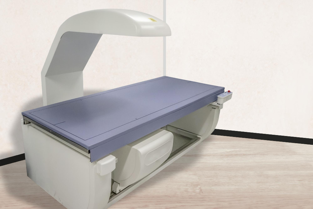
What Happens During a DEXA scan?
- A DEXA scan will be performed by a radiologist.
- The patient will be asked to lie down on an x-ray table and asked to stay still.
- A scanning machine will pass above the patient’s body passing a low dose of x-ray beams.
- Some of the x-rays will be absorbed by the soft tissues.
- An x-ray detector will measure the x-rays that pass through; using this information, the density of bones will be determined.
More About the Scan
- There will be no discomfort of any kind during the scan.
- It is a completely safe procedure.
- However, pregnant women (or women who suspect pregnancy) should not undergo the scan.
- It is a very quick diagnostic procedure the duration of which takes only around 20 minutes.
- Calcium supplements should be stopped 24 hours ahead of the scan.
- Comfortable clothing without any zippers or metal components is best.
- Jewelry cannot be worn during the scan.
Results of the Scan
The patient’s scan results will be compared with the bone density of a healthy young adult as well as with the bone density of a healthy adult who is the same age, sex and ethnicity as the patient. After comparison, two values are obtained. This is called the standard deviation (SD). Two scores are calculated:
- T-score: Difference in bone density of patient and healthy young adult.
- Z-score: Difference in bone density of patient and healthy adult with the same parameters as the patient. Z score is used for determining the bone density of children and adults till 30 years of age.
The T & Z scores are interpreted as follows:
- T score > -1: Normal
- T score falls between -1 and -2.5: Mild reduction in bone density
- T score <= -2.5: Osteoporosis
- Z score < -2: Bone density is low
Frequency of Scan
Doctors recommend that a DEXA scan be done once every two years. However, this is subject to change depending on the patient; factors like age, gender, fracture risk, etc. will all be taken into account to determine the frequency of the scan. The doctor will then give a comprehensive plan to safeguard bone health.
Accuracy of Scan
DEXA scan results are considered to be highly accurate and are preferred by doctors to measure bone density. However, in case of special circumstances, (for example, when a patient has had previous spinal surgery or has spondylosis) the accuracy of the scan may be affected.
In conclusion, we can say that a DEXA scan is an effective tool for doctors to diagnose osteoporosis and plan for fracture prevention. As the body is exposed to only very low doses of radiations, the gains offset the risk.